Dendrimer nanosystems for adaptive tumor-assisted drug delivery via extracellular vesicle hijacking
Yifan Jiang, Zhenbin Lyu, Brigino Ralahy, Juan Liu, Tom Roussel, Ling Ding, Jingjie Tang, Artemis Kosta, Suzanne Giorgio, Richard Tomasini, Xing-Jie Liang, Nelson Dusetti, Juan Iovanna, Ling Peng
PNAS, 2023, Vol. 120, No. 7, e2215308120
https://doi.org/10.1073/pnas.2215308120
Cancer is one of the leading causes of death in the world, and remains a difficult disease to treat because of its intrinsic heterogeneity and dynamic evolution. Nanosystems (DDSs) that can overcome tumor heterogeneity and dynamic evolution, yet delivering anticancer drug deep in tumor tissue are desperately demanded for cancer treatment. However, such systems are difficult and challenging to develop. We report a drug delivery nanosystem based on self-assembling dendrimer nanomicelles (ADNMs) for deep tumor penetration and effective drug delivery via in situ tumor-secreted extracellular vesicles (EVs), an endogenous transport system that evolves with the parental cells in the tumor microenvironment, hence overcoming tumor heterogeneity and dynamic evolution, leading to excellent anticancer activity. Specifically, these dendrimer nanomicelles, upon arrival at a tumor, had their drug payload repackaged by the cells into EVs. Those were further transported and internalized by other cells for delivery “in relay”, deep into the tumor tissue improving drug delivery efficacy and cancer cell killing (Figure 1). Using pancreatic and colorectal cancer-derived 2D, 3D, and xenograft models, we demonstrated that the in situ-generated EVs mediated intercellular delivery (Figure 2), propagating drug delivery from cell to cell deep into the tumor, leading to excellent anticancer potency yet reducing adverse effects (Figure 3). These promising results highlight the remarkable potential of this drug delivery system for cancer treatment and provide a proof of concept for the development of new therapeutic modalities for adaptive drug delivery. This study also provides a new perspective on exploiting the intrinsic features of tumor alongside dendrimer supramolecular chemistry to develop smart and effective DDSs to overcome intra-tumoral microenvironment and heterogeneity as their evolutive nature, thereby improving cancer therapy.
This collaborative study involved the Centre Interdisciplinaire de Nanoscience de Marseille (CINaM), the Centre de Recherche en Cancérologie de Marseille (CRCM) and the National Center for Nanoscience and Technology (NCNST) of China.
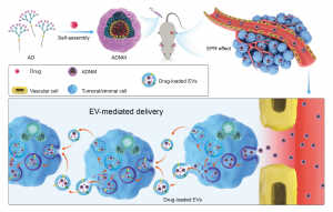
Figure 1: Amphiphilic dendrimer nanomicelles (ADNMs) encapsulate the anticancer drug and induce tumor-assisted drug delivery via extracellular vesicle (EV)-mediated intercellular transport. The amphiphilic dendrimer (AD) encapsulates the anticancer drug and forms nanomicelles (ADNMs) which reach the tumor lesion via the enhanced permeability and retention (EPR) effect. There they induce in situ tumor-assisted drug delivery for deep tumor penetration via EV-mediated intercellular transport. This EV-mediated delivery process involves: (1) internalization of ADNMs inside cells within the tumor tissue; (2) repackaging of ADNM payload into EVs; (3) intercellular transport of the generated EVs; (4) internalization of the generated EVs by the recipient cell.
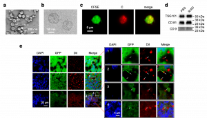
Figure 2. ADNM induced EV payload-packaging and cellular uptake. EVs, generated by cells upon treatment with ADNMs (R/AD, Cy3/R/AD and Dil/R/AD), were characterized using TEM (a), cryogenic electron microscopy (Cryo-EM) (b), fluorescent microscopy (c) and EV using western blotting (d). e, Confocal images of cryo-sectioned tumor tissues from HCT-8GFP xenograft mice treated with ADNM (DiI/R/AD) show the process of EV-mediated delivery in the tumor. The DiI fluorescence appeared in EVs derived from HCT-8GFP tumor (arrows). The hollow-donut shape of red DiI signal with green GFP filling highlights the HCT-8GFP-derived EVs with the DiI-labelled phospholipid bilayer and the HCT-8GFP-derived contents inside. Box 1, a DiI-loaded EV (arrow) located within the intercellular space; Box 2–4, the DiI-loaded EVs mediated intercellular transport within the tumor. Box 2, DiI-loaded EVs adhered onto the cell surface; Box 3, a DiI-loaded EV entering into a cell; Box 4, a DiI-loaded EV inside a cell. The tumor tissues were collected 24 hours after intravenous injection of DiI/R/AD in HCT-8GFP xenografts. AD: amphiphilic dendrimer; C: Cy3; Dil: fluorescent dye; R: rapamycin.
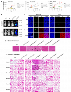
Figure 3. ADNMs were effective for inhibiting tumor growth, reducing drug toxicity and preventing tumor metastasis via specific accumulation and deep penetration through EVs in tumor. a, Tumor growth curves of the patient-derived pancreatic cancer xenografts PDAC087T and PDAC074T, and the colorectal cancer HCT-8 xenografts in mice upon treatment with ADNM carrying either doxorubicin (DOX) or rapamycin R, respectively. Mice treated with PBS buffer, drug alone or dendrimer alone were used as the controls. Data are presented as the mean ± s.e.m. The statistical significance was calculated by two-way ANOVA with a Tukey’s multiple comparisons test. n = 6 mice for all groups. *P < 0.05, **P < 0.01, ***P < 0.001, ****P < 0.0001. b, Accumulation of R/AD in tumors in the PDAC074T and HCT-8 xenograft mice was analyzed using fluorescent imaging with ADNM carrying both rapamycin and the near-infrared fluorescent dye DiR (DiR/R/AD). The in vivo fluorescence images were acquired 48 hours after intravenous administration of DiR/R/AD in PDAC074T (upper panel) and HCT-8 (lower panel) xenografts. Mice treated with PBS, free DiR, and a simple mixture of DiR, rapamycin and AD (DiR+R+AD), were used as controls. Arrows point to the tumor locations. c, Confocal images of tumor tissues show a deep intratumoral penetration of R/AD, using ADNM loaded with both rapamycin and the fluorescent dye DiI (DiI/R/AD). Mice treated with PBS, DiI alone, or a simple mixture of DiI, rapamycin and AD (DiI+R+AD), were used as controls. Tumors were harvested from PDAC074T (left) and HCT-8 (right) xenografts 24 hours post-intravenous administration, then cryosectioned and imaged by tracing DiI fluorescence (red). Blood vessels were labelled with DyLight488-labelled Lycopersicon esculentum lectin (green), and nuclei with DAPI (blue). d, HE stain of heart tissue issued from PDAC087T xenografts treated with PBS, AD, DOX and DOX/AD (n = 6). Treatment with DOX/AD prevented DOX-induced hyperemia and myocardial fiber breakage (arrows) in heart. e, HPS stain of lung tissues issued from HCT-8 xenografts treated with PBS, AD, R and R/AD (n = 6). Treatment with R/AD prevented the rapamycin-induced lung metastasis (T represents the tumor). Lung metastases were observed in mice treated with R, but not in those treated with PBS, dendrimer alone (AD) or the R/AD. Lung tissue was collected from mice at the end of treatment.