Mechanisms of splenic filtration
Principal investigators: A. Viallat, E. Helfer, A. Charrier
PhD students : Alexis Moreau (2017-2021), Anagha Surendranath (2021-2022)
Postdoc : François Yaya (2021-2023), Cécile Iss (2016-2017), Scott Atwell (2015-2017), Priya Gambhire (2015-2016)
Collaborators: Zhangli peng (Univ . Notre Dame, Notre Dame & UIC, Chicago, U. S. A.), Pierre Buffet (BIGR, Paris)
Funding: ANR SpleenMark (2021-2024), AMIDEX RedPath (2015-2017), CENTURI PhD grant (2021-2022), PhD doctoral school fellowship (2017-2020),
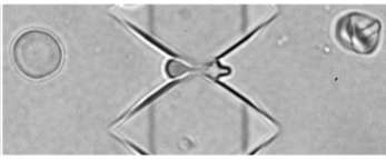
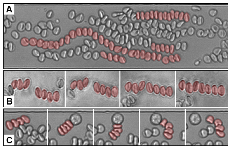
In the bloodstream, RBCs not only circulate in vessels and capillaries, but also pass through the spleen, an organ whose main function is to filter blood. There, RBCs must pass through narrow slits which are sub-micrometric in width and a few µm in height and length, thus undergoing extreme deformations. We developed a microfluidic device containing submicron slits mimicking the splenic slits to study the stringent mechanical fitness test that red blood cells pass in the spleen.
Initial proof-of-concept experiments were carried out by passing RBCs from healthy donors and patients with blood diseases that rigidify the RBCs. We observed previously unreported behaviours which we linked to the mechanical specificities of healthy and diseased RBCs. This device thus enables us to discriminate GR behaviours according to their mechanical properties.
We quantitatively assessed the passage of healthy RBCs through these biomimetic slits and observed that they can pass through 0.3-µm wide slits at body temperature. Our experimental results, combined with numerical simulations, showed that circulating RBCs have enough surface area excess to deform into 2 spheres connected by a tether and that those with the lowest surface area locally unfold their spectrin cytoskeleton inside the narrower slits.
Physical mechanisms of red blood cell splenic filtration. A. Moreau, F. Yaya, H. Lu, A. Surendranath, A. Charrier, B. Dehapiot, E. Helfer, A. Viallat, Z. Peng. Proceedings of the National Academy of Sciences 120, e2300095120 (2023). https://doi.org/10.1073/pnas.2300095120
Reply to Kaestner et al.: Activation of PIEZO1 is not significant for the passage of red blood cells through biomimetic splenic slits. A. Moreau, F. Yaya, H. Lu, A. Surendranath, A. Charrier, B. Dehapiot, E. Helfer, A. Viallat, Z. Peng. Proceedings of the National Academy of Sciences 122, e2411469121 (2025). https://doi.org/10.1073/pnas.2411469121
Analytical theory for a droplet squeezing through a circular pore in creeping flows under constant pressures. Z. Tang, F. Yaya, E. Sun, L. Shah, J. Xu, A. Viallat, E. Helfer, Z. Peng. Physics of Fluids 35, 082016 (2023). https://doi.org/10.1063/5.0156349
Transit time theory for a droplet passing through a slit in pressure-driven low Reynolds-number flows. S. W. Borbas, K. Shen, C. Ji, A. Viallat, E. Helfer#, Z. Peng#. Micromachines 14, 2040 (2023). https://doi.org/10.3390/mi14112040
High aspect ratio sub-micrometer channels using wet etching: Application to the dynamics of red blood cell transiting through biomimetic splenic slits. P. Gambhire, S. Atwell, C. Iss, F. Bedu, I. Ozerov, C. Badens, E. Helfer, A. Viallat, A. Charrier. Small 13, 1700967 (2017). https://doi.org/10.1002/smll.201700967
Collective dynamics of red blood cells under confined 2D-flow
Principal investigators: E. Helfer, A. Viallat, A. Charrier
Postdoc : Marie Poulain-Zarcos (2022-2023), Cécile Iss (2016-2017)
PhD student : Alexis Moreau (2017-2018)
Collaborators: Emilie Franceschini (LMA, Marseille, France) ; Laurence Bergougnoux (IUSTI, Marseille, France) ; Simon Mendez (IMAG, Montpellier, France)
Funding: CENTURI postdoctoral fellowship (2022-2023), PhD doctoral school fellowship (2017-2020), AMIDEX RedPath (2015-2017)
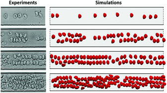
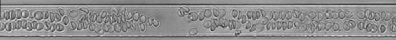
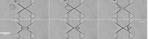
Emulsions/suspensions are widely used in many fields, both fundamental and applied. The description of the structuration of these emulsions under flow is incomplete, notably due to the lack of confrontation with experimental data caused by the lack of controlled deformable particles. We tackled this purely physical problem by using red blood cells as model deformable particles and studied their behavior under confined 2D flow. By combining our microfluidic experiments with numerical simulations, we demonstrated the effect of lateral confinement and the crucial role of deformability on the lateral organization of red blood cells into parallel lines along the flow.
Self-organization of red blood cell suspensions under confined 2D flows. C. Iss, D. Midou, A. Moreau, D. Held, A. Charrier, S. Mendez, A. Viallat#, E. Helfer#. Soft Matter 15, 2971 (2019). https://doi.org/10.1039/c8sm02571a
Mechanical adaptation of monocytes in model pulmonary capillary networks
Principal investigators: A. Viallat, E. Helfer
PhD student : Jules Dupire (2010-2012)
Collaborator: Pierre-Henri Puech (LAI, Marseille)
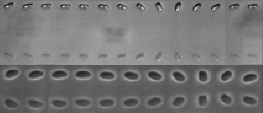
Correct circulation of white blood cells (WBCs) in the pulmonary vascular bed is crucial for an effective immune response. In this ramified vascular network, WBCs must deform strongly to pass through the narrowest capillaries and bifurcations. Although this process is known to depend on the mechanical properties of cells, it is still poorly understood due to the lack of a comprehensive model of cell mechanics and physiologically relevant experiments. Here, using an internal microfluidic device mimicking the pulmonary capillary bed, we showed that the dynamics of THP1 monocytes evolve along successive capillary-like channels, from a slow non-stationary motion with jumps to an efficient fast and smooth motion. We used actin cytoskeleton drugs to modify trafficking dynamics. This led us to propose a simple mechanical model showing that a very finely tuned cortical tension combined with high cell viscosity governs rapid transit through the network while preserving cell integrity. Finally, we demonstrated that cortical tension controls steady-state cell velocity via viscous friction between the cell and the channel walls.
Mechanical adaptation of monocytes in model lung capillary networks. J. Dupire, P.-H. Puech, E. Helfer, A. Viallat. Proceedings of the National Academy of Sciences 117, 14798 (2020). https://doi.org/10.1073/pnas.1919984117